Authors: Dr. Raphael Monte Alto, Dra. Helena Klemba, Dra. Ariane Vaz Storrer, Dra. Francieli Suntak, Dra. Luany Halaiko, Jucimara Klemba and TPD Miguel Abrão
Initial Evaluation
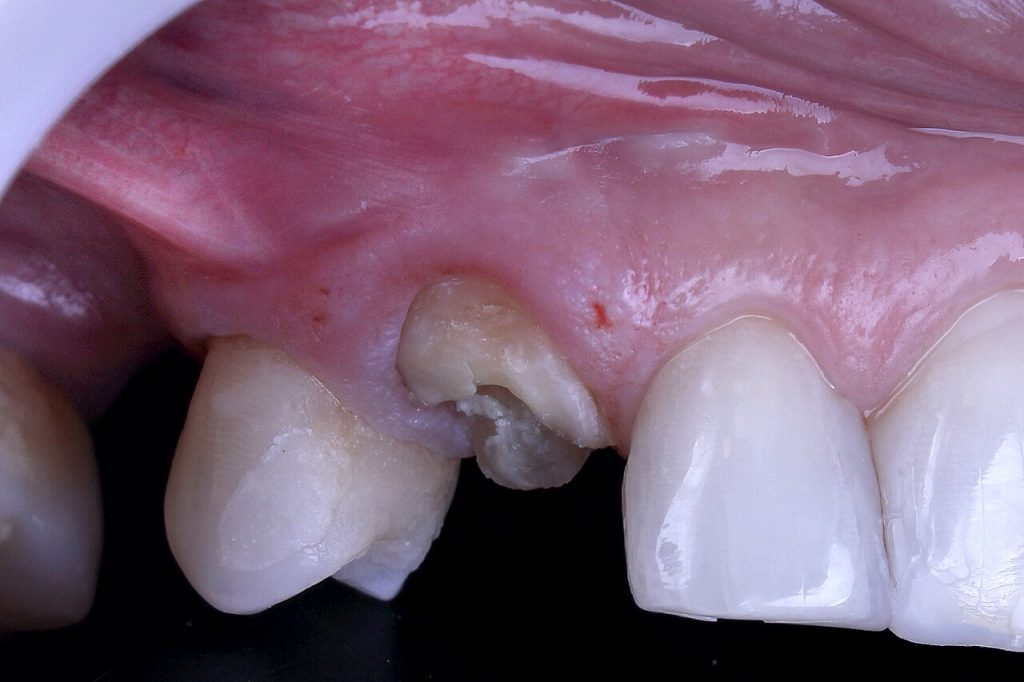
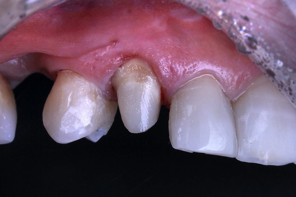
After anamnesis, clinical and radiographic examination, a deficient provisional cemented acrylic resin full crown was observed. The endodontic treatment was adequate with no pain symptoms or periapical changes.
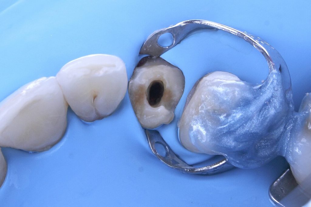
After crown removal, the presence of little remaining coronal was observed, so the reconstruction was planned through an anatomic fiberglass post and future full ceramic crown.
Treatment
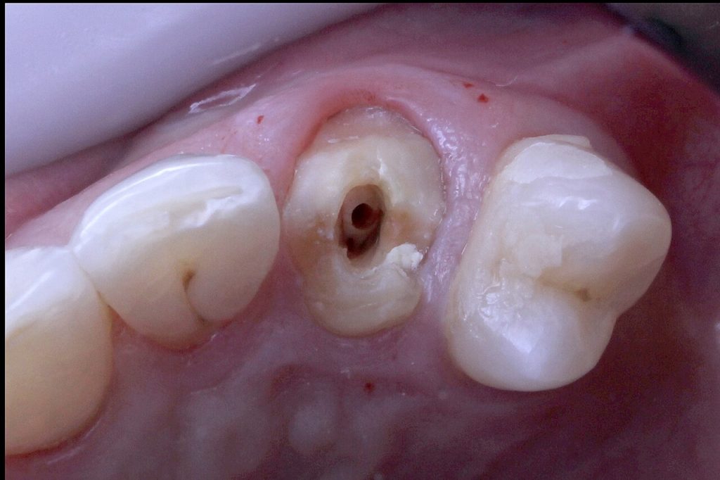
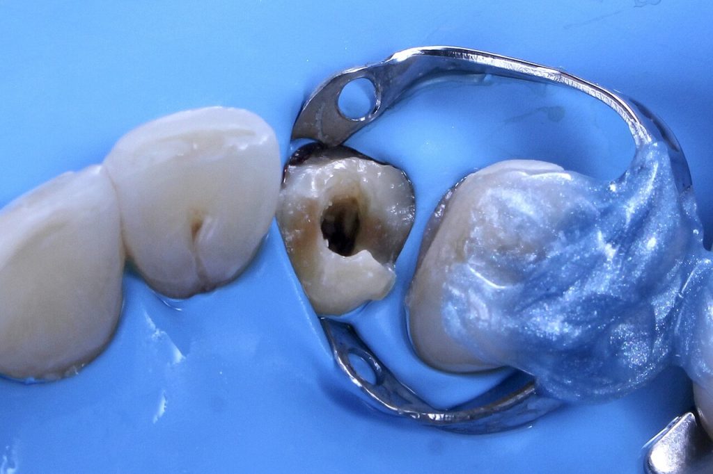
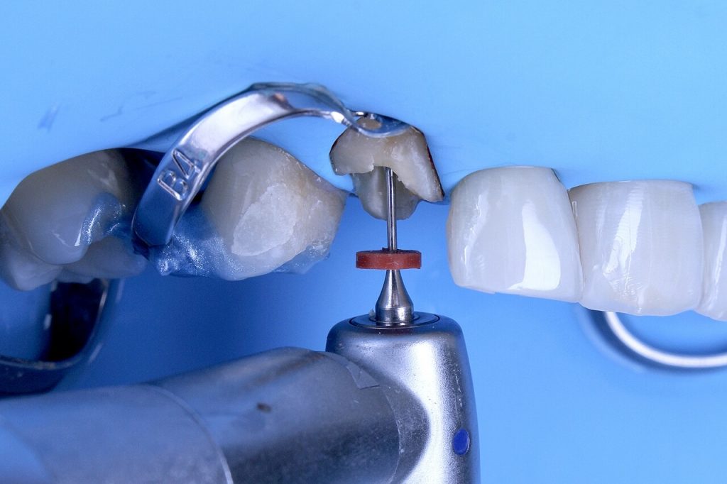
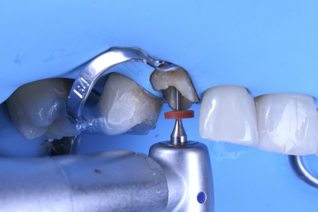
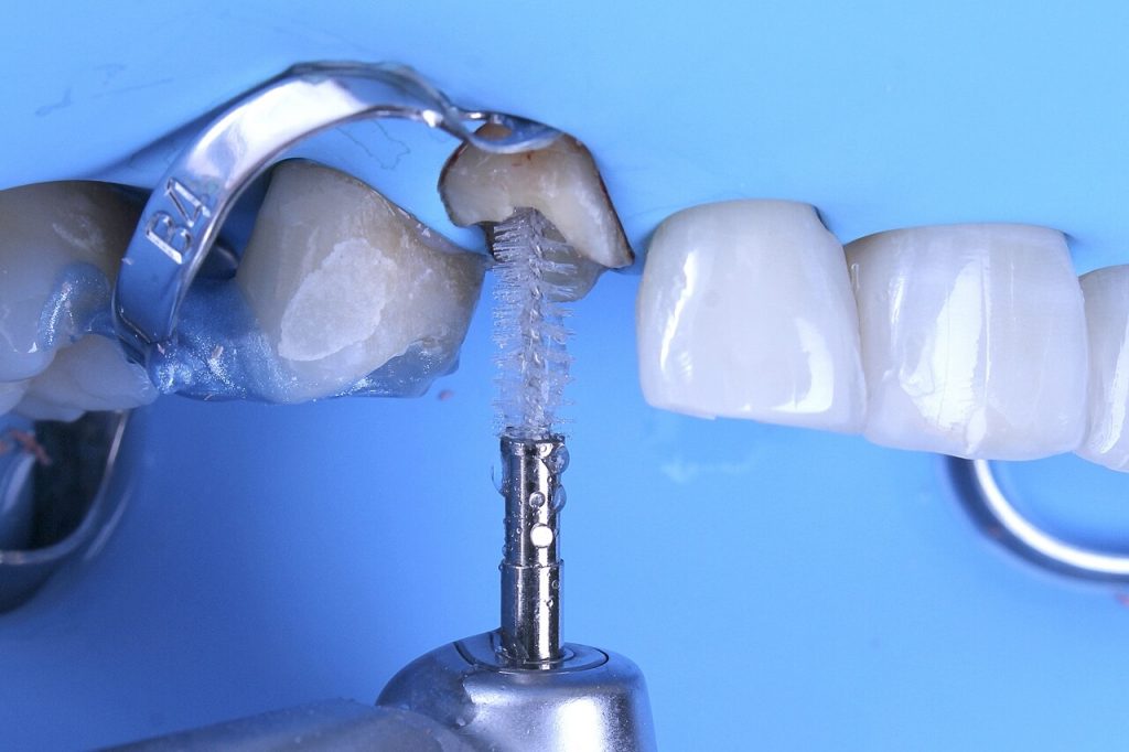
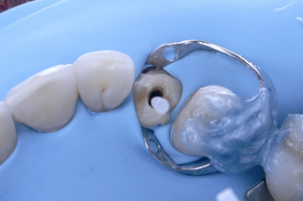
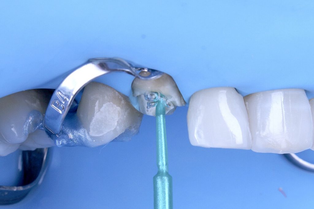
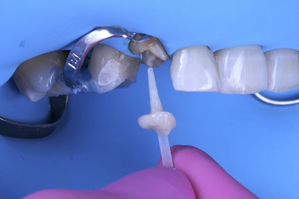
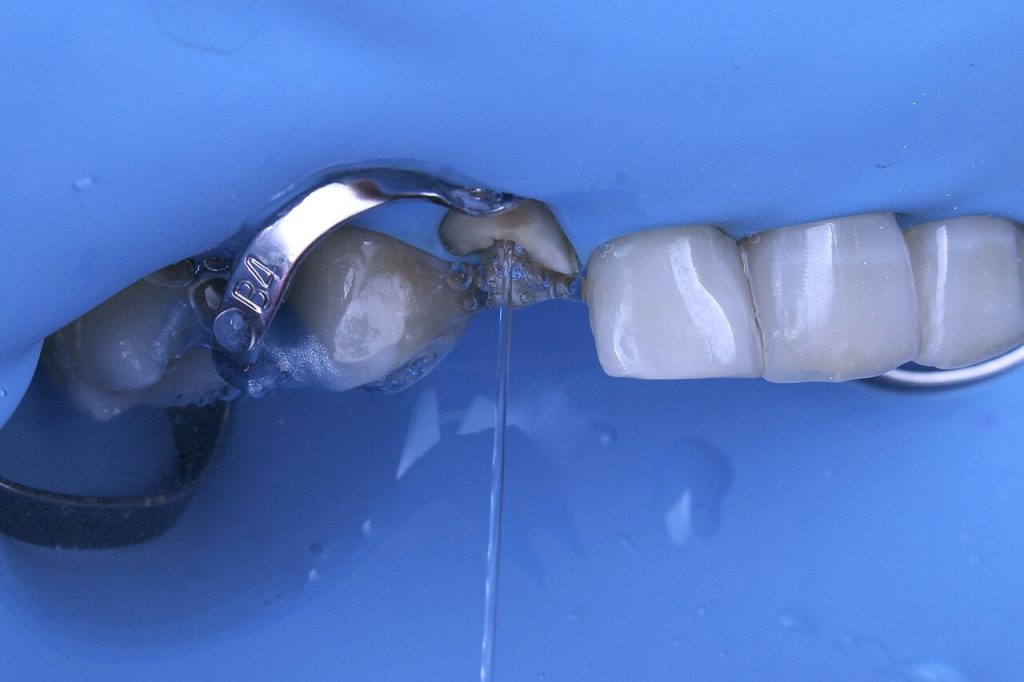
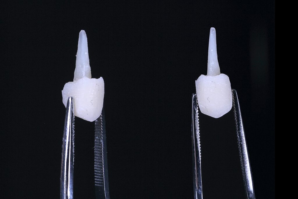
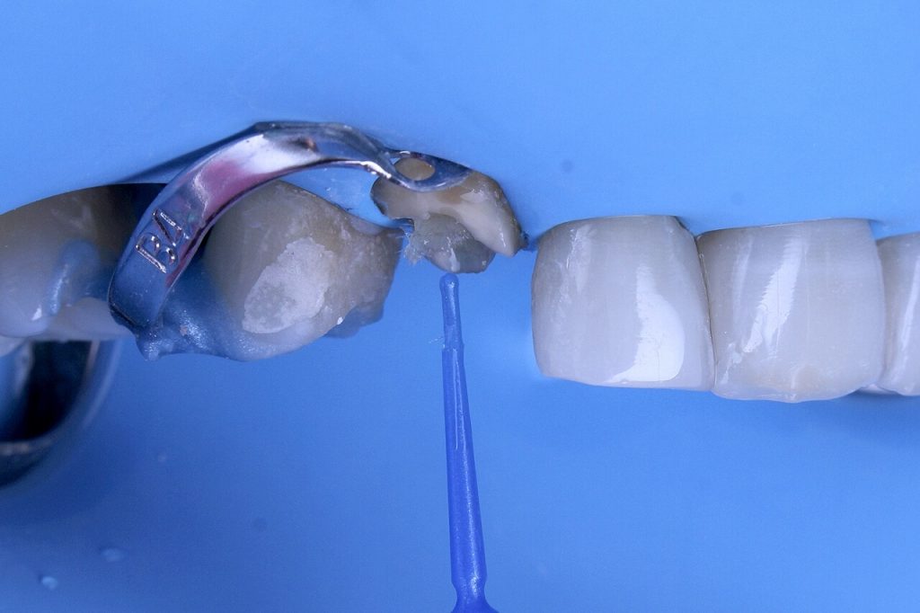
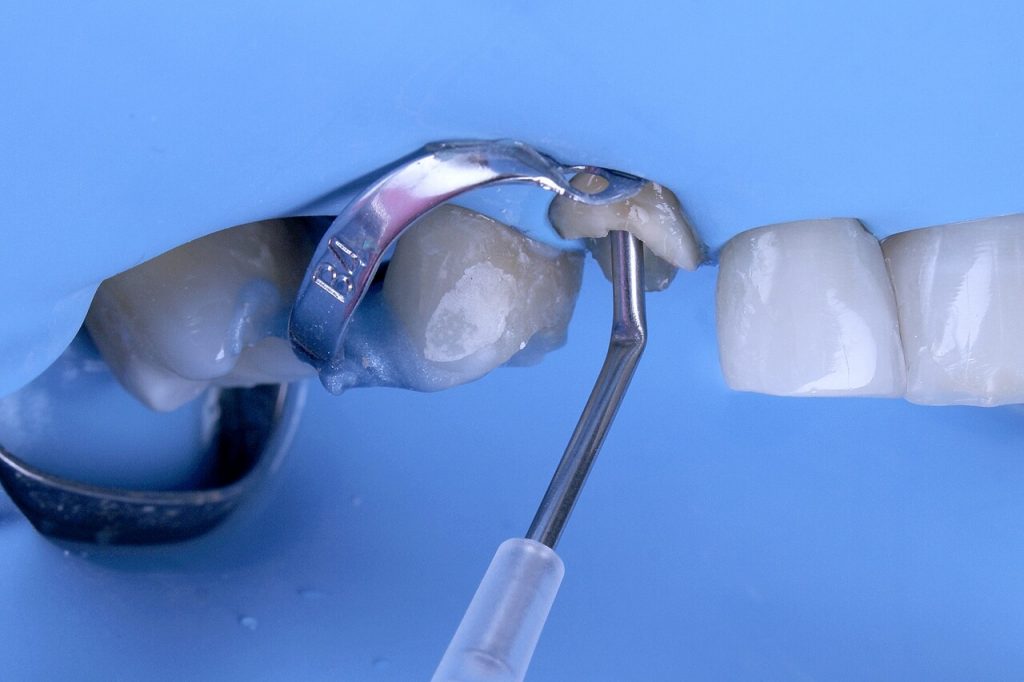
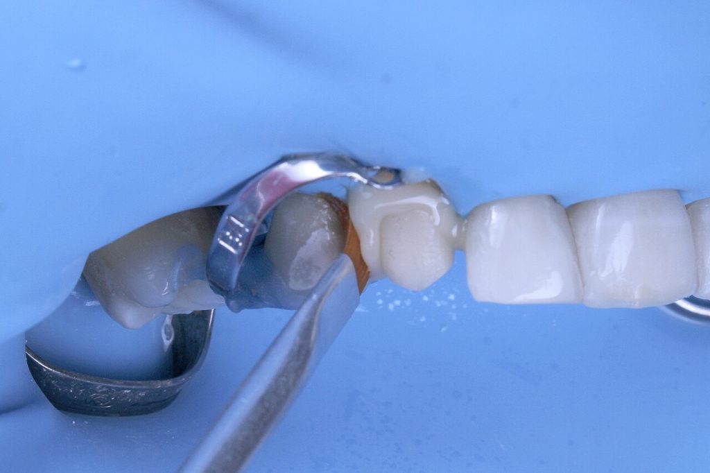
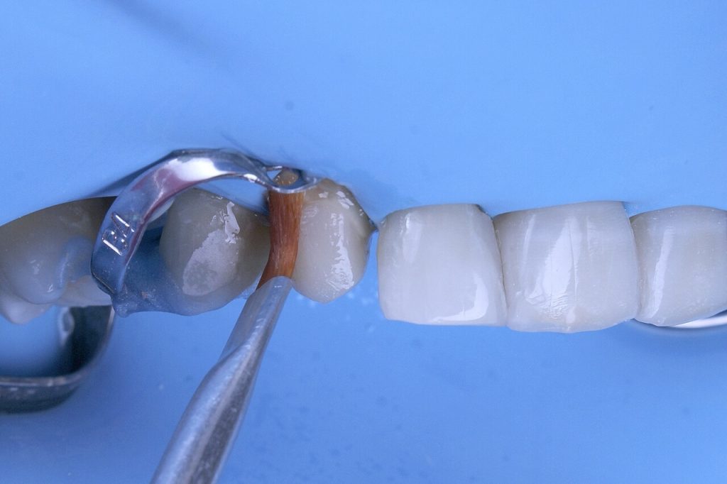
The resin restoration was removed and the coronal remnant was evaluated. After performing odonometry using a size 2 large bur, the gutta-percha was removed, keeping 5 mm in the apical region. The canal was cleaned with an intracanal prophylaxis brush to remove gutta-percha and endodontic cement remnants. After cleaning the canal, the Whitepost System DC 1 post was tried in. The choice of a post with a smaller diameter than the canal aims to anatomize the post with composite resin. This procedure, in addition to allowing better frictional retention with the canal, allows the post to be positioned centrally to the composite resin core.
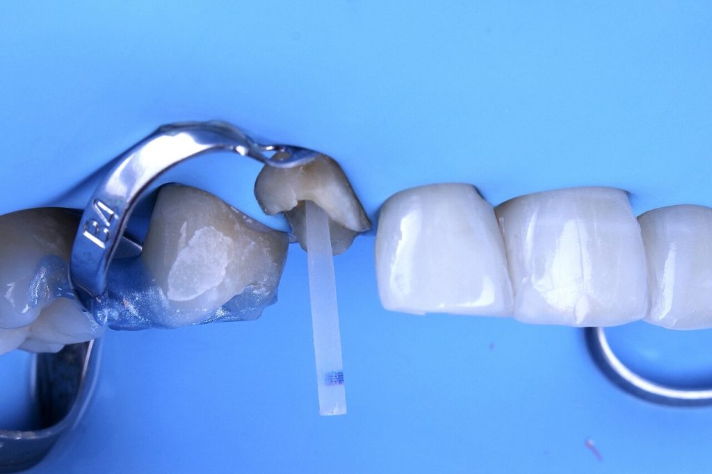
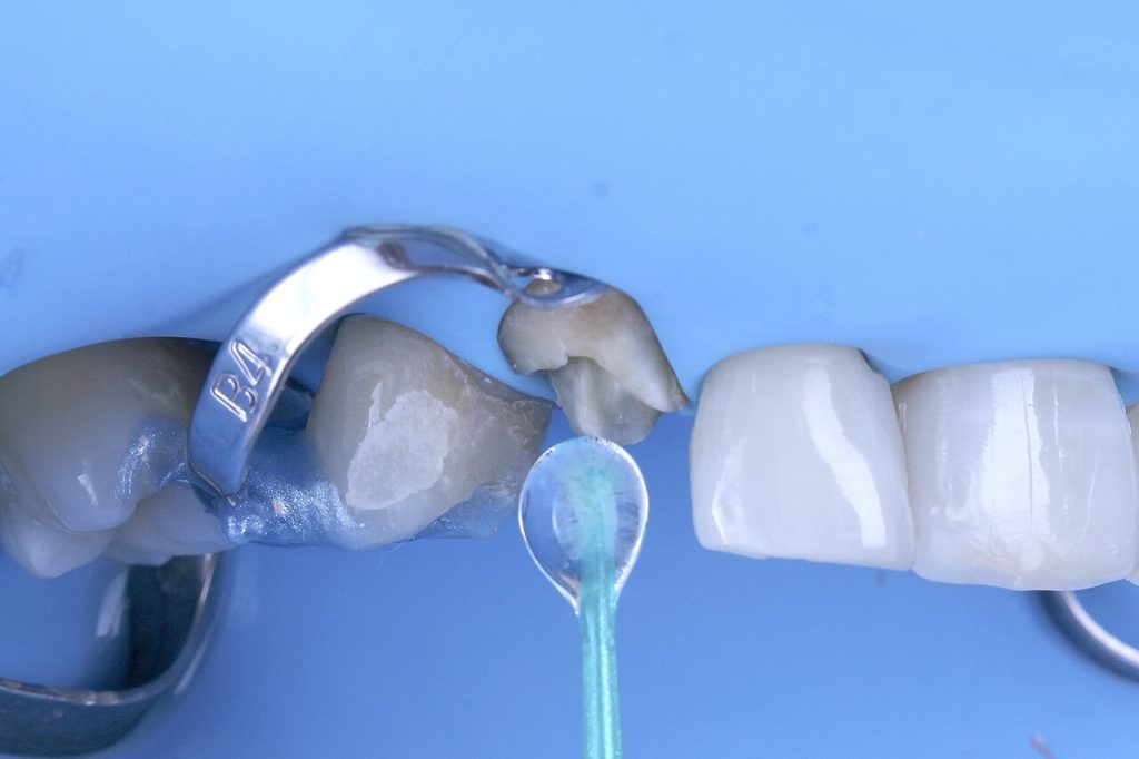
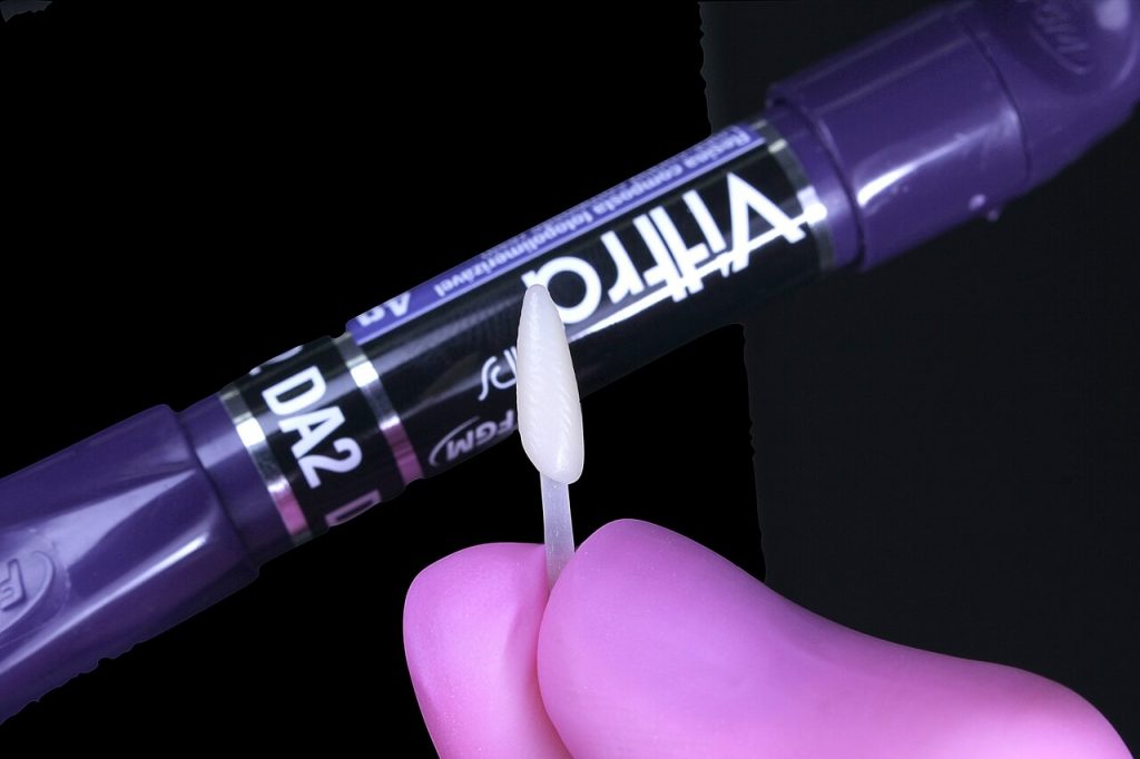
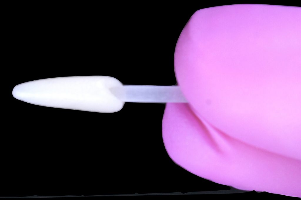
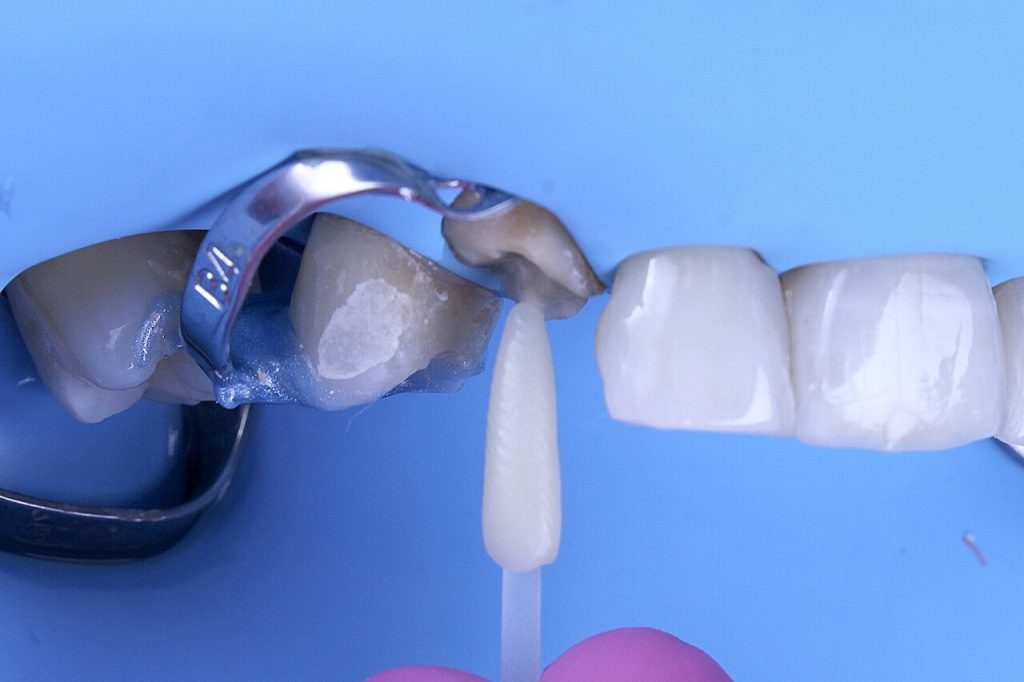
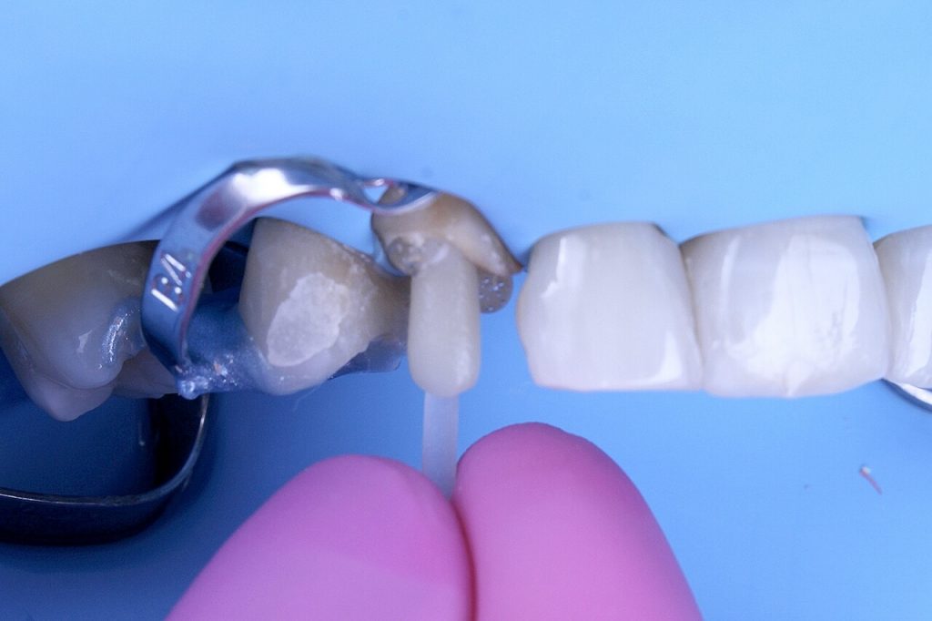
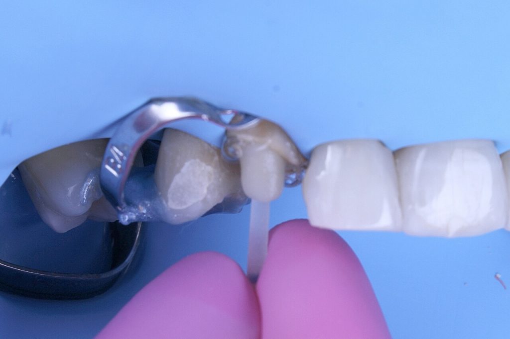
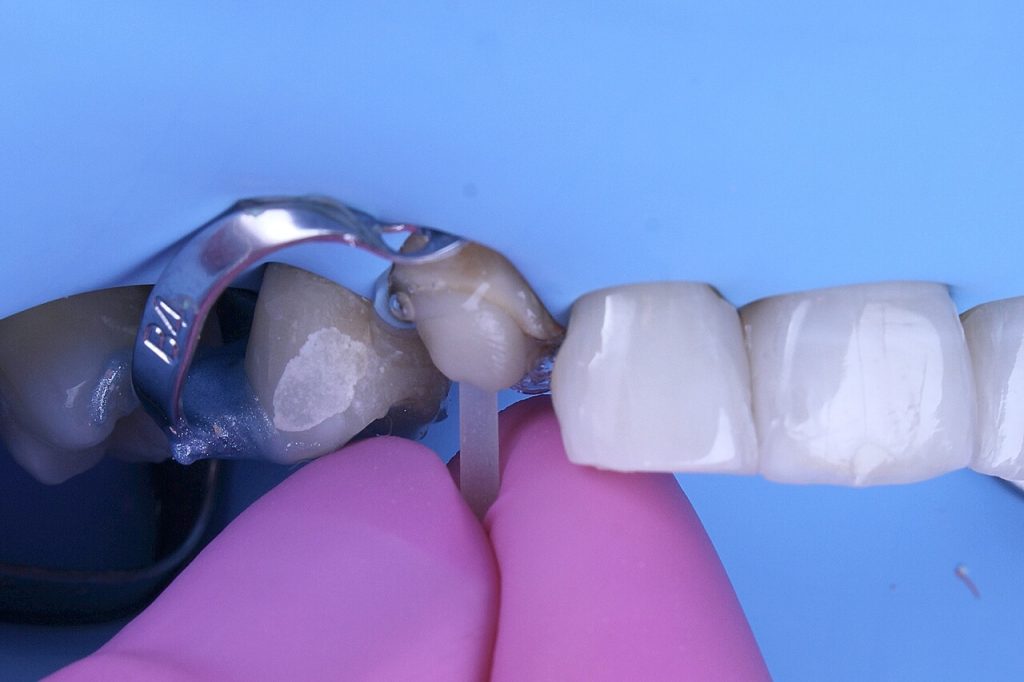
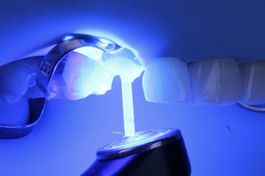
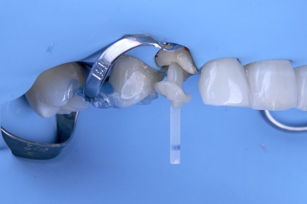
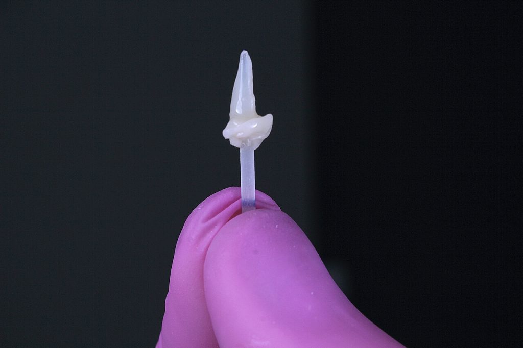
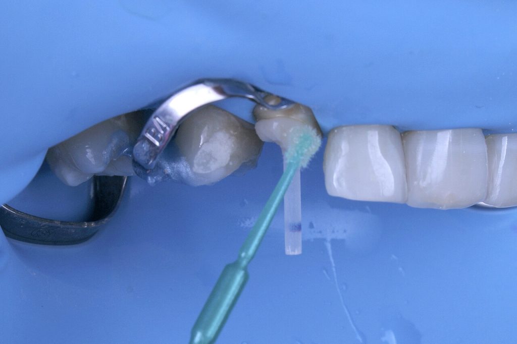
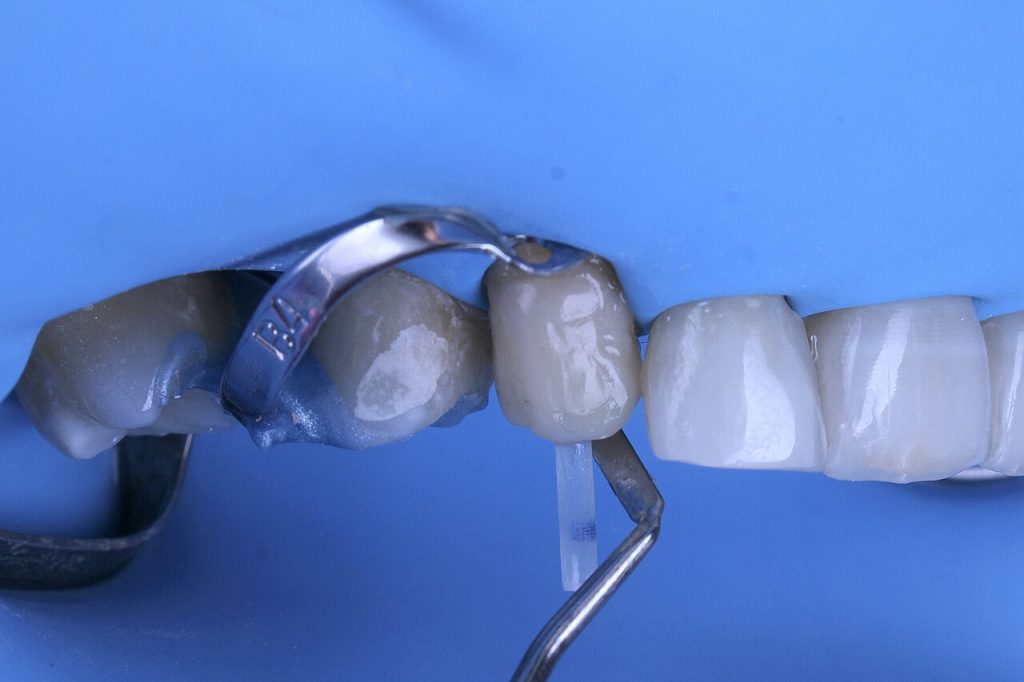
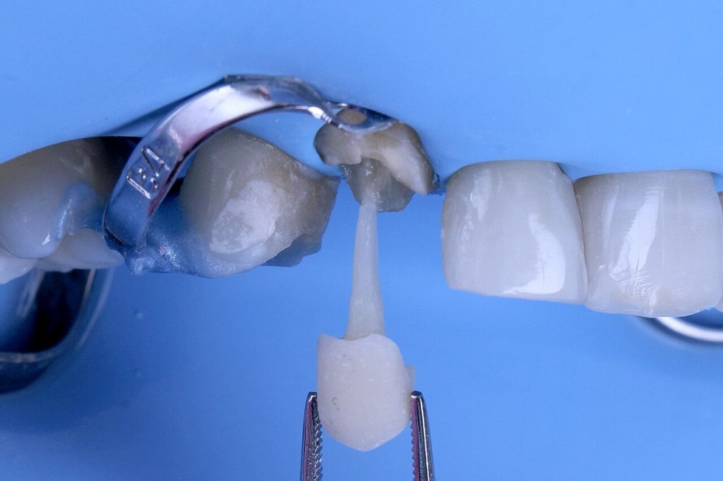
To prevent the composite resin from being retained in the canal, it was isolated with a water-based gel. The post was cleaned with gauze and alcohol, and an adhesive was applied to its surface. A cone was modeled with a Vittra APS (DA2) dentin composite resin over the apical portion of the fiberglass post, with dimensions compatible with the canal. The assembly was then slowly taken so that the composite resin would copy the shape of the canal. After the total insertion of the post in its odontology, the cervical composite resin was slightly condensed for perfect adaptation in the cervical region. A photoactivation of 5s was performed with a photoactivator, then about 2mm of the post was removed, returned to the same position and 5s more light was applied. This procedure is performed until the post comes out almost completely polymerized.
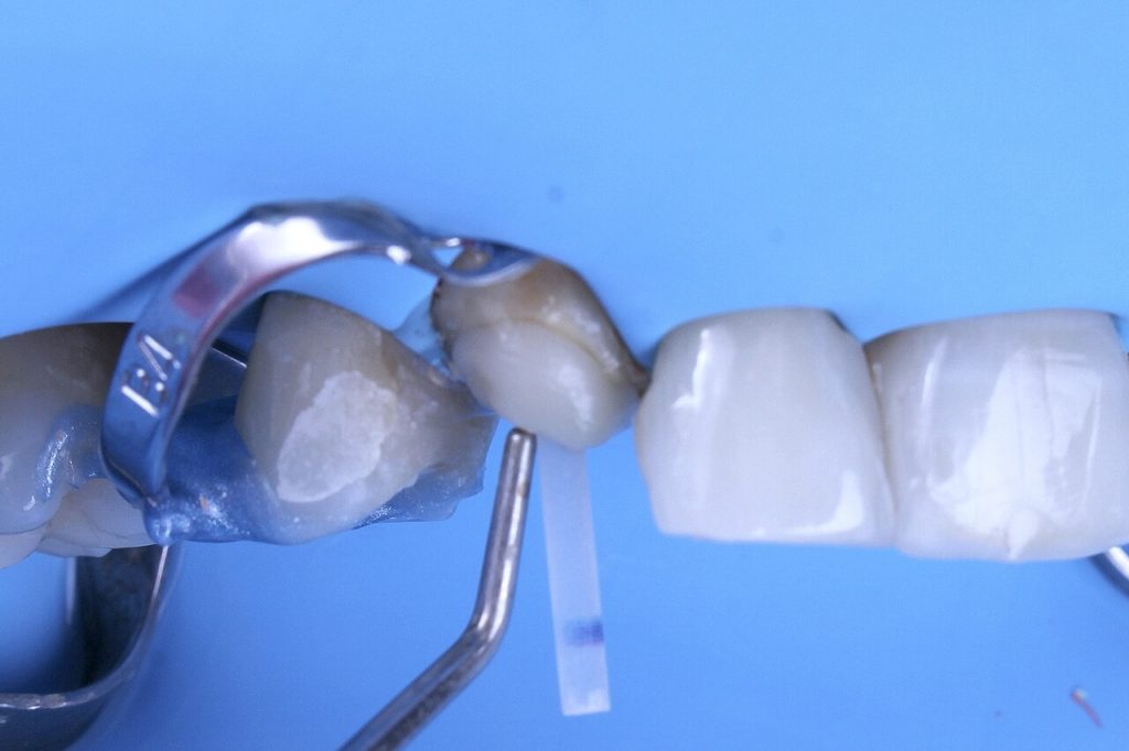
Before completely removing the pin, a mark is made to identify the buccal surface.
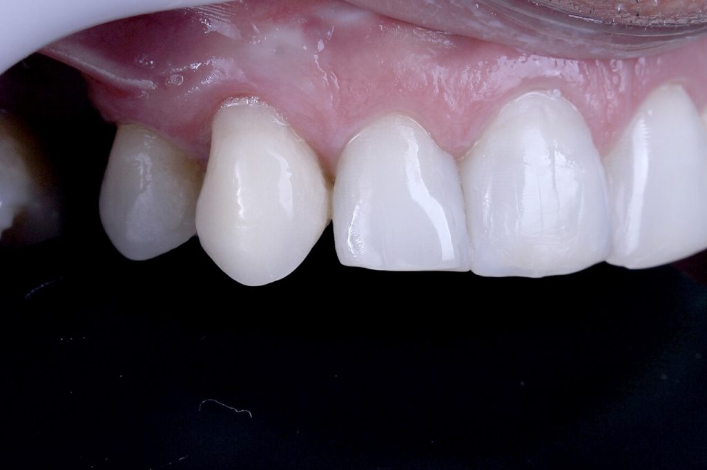
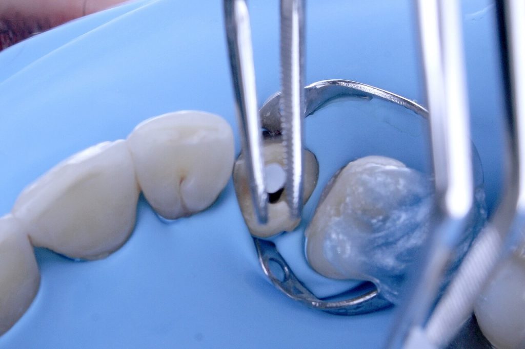
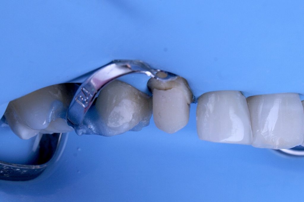
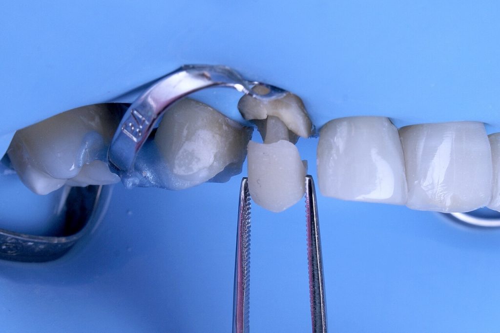
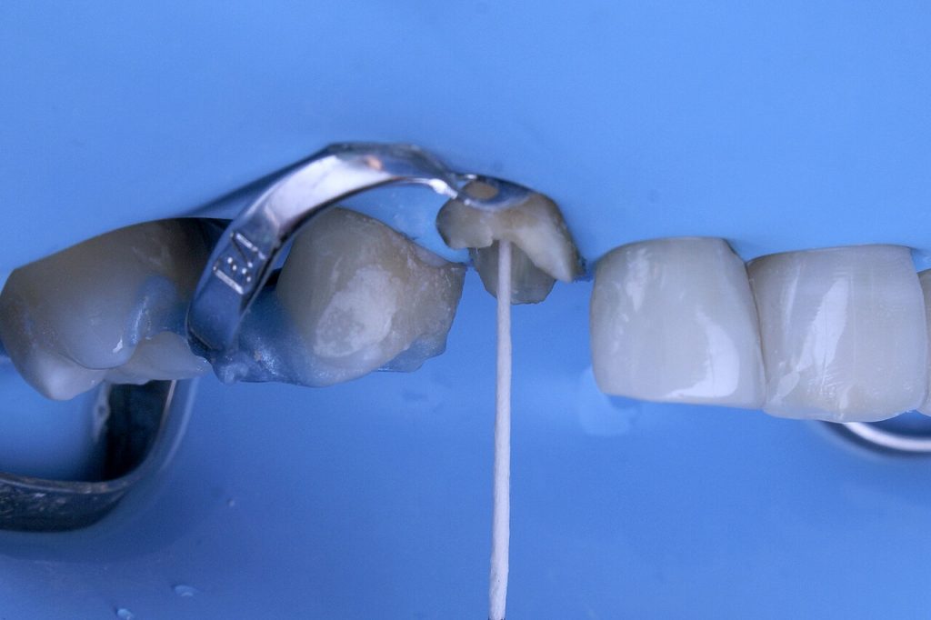
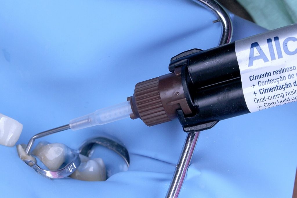
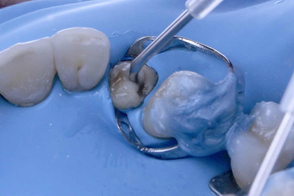
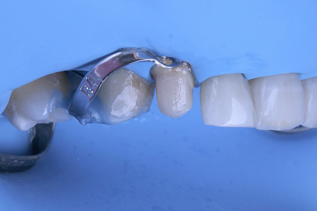
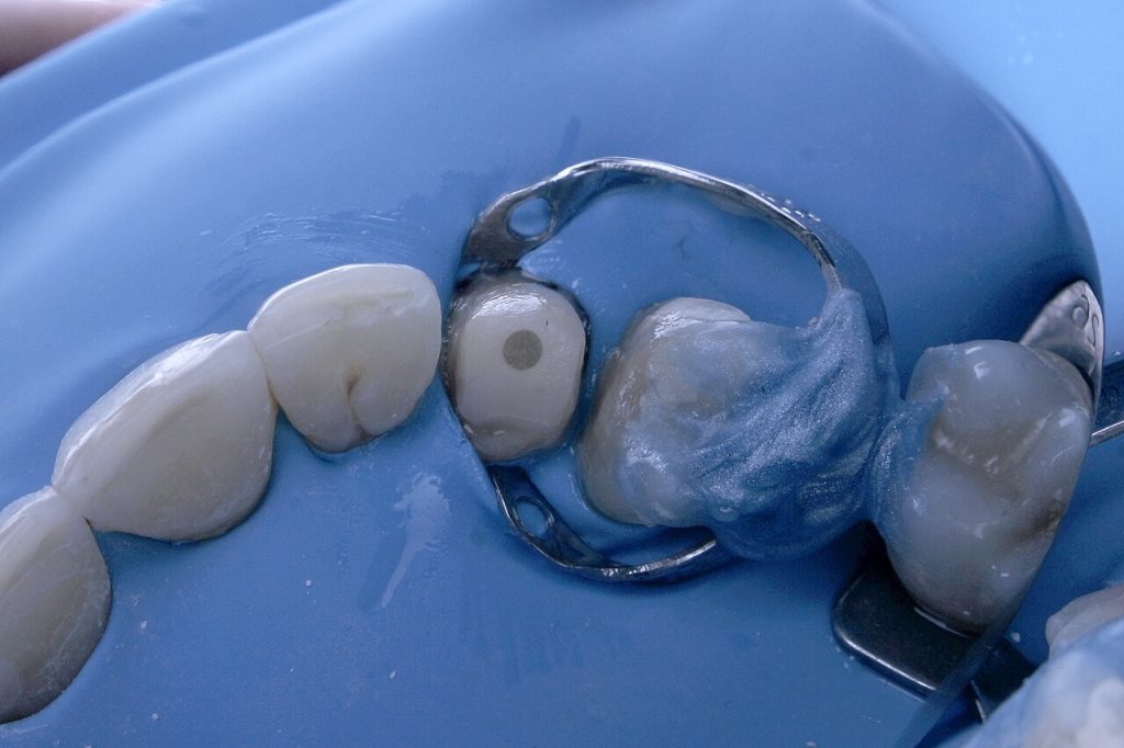
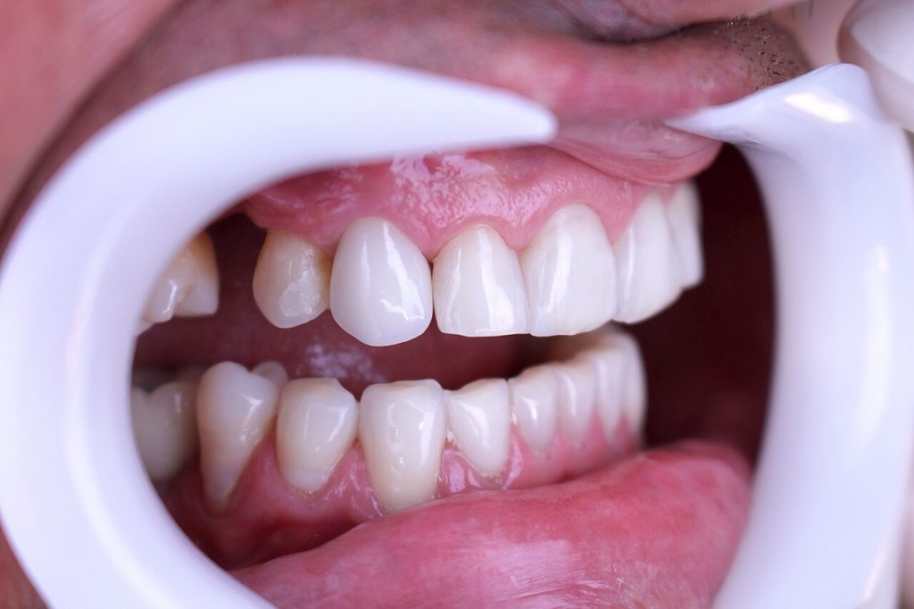
Complementary polymerization is performed with the resin post set outside the mouth. The canal was washed, the post was reinserted, and the coronal portion was filled with the same composite resin. With the assembly in position, the preparation for a full crown is performed, and immediately after, the assembly is removed with a hemostatic clamp. For final cementation, the canal was thoroughly washed, dried with paper cones, and the post cleaned with a gauze and alcohol. Ambar Universal APS adhesive was applied to the canal and Allcem Core cement was used for final cementation. After removing the isolation, the preparation was finalized and a mold was taken. The mold was sent to the Singulares laboratory (TPD Miguel Abrão) where a disilicate crown was made. The crown was cemented with Allcem Veneer APS and Ambar Universal APS.
Step-by-step
