Treatment of a fracture on an upper central incisor with rehabilitation through immediate implant carried out with a virtual surgical guide: a clinical case report

Authors: Prof. Augusto Bessa and Prof. Íris Sol

Initial evaluation

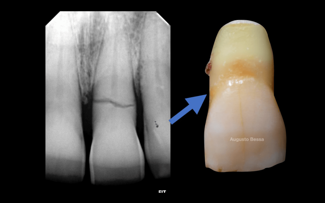
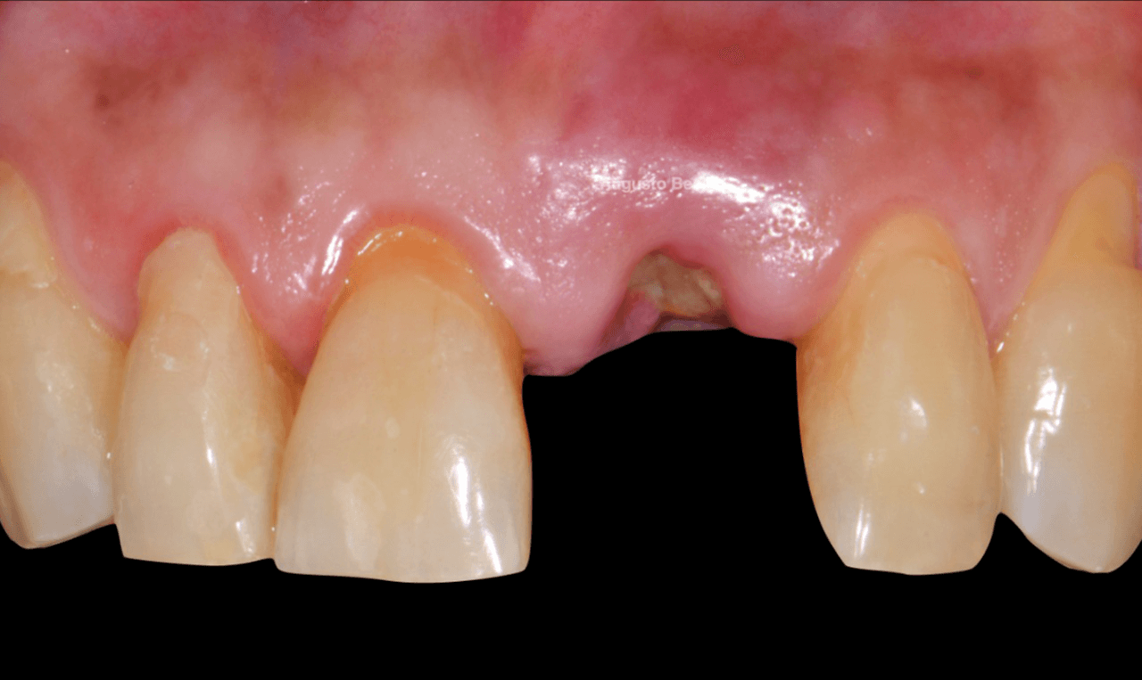
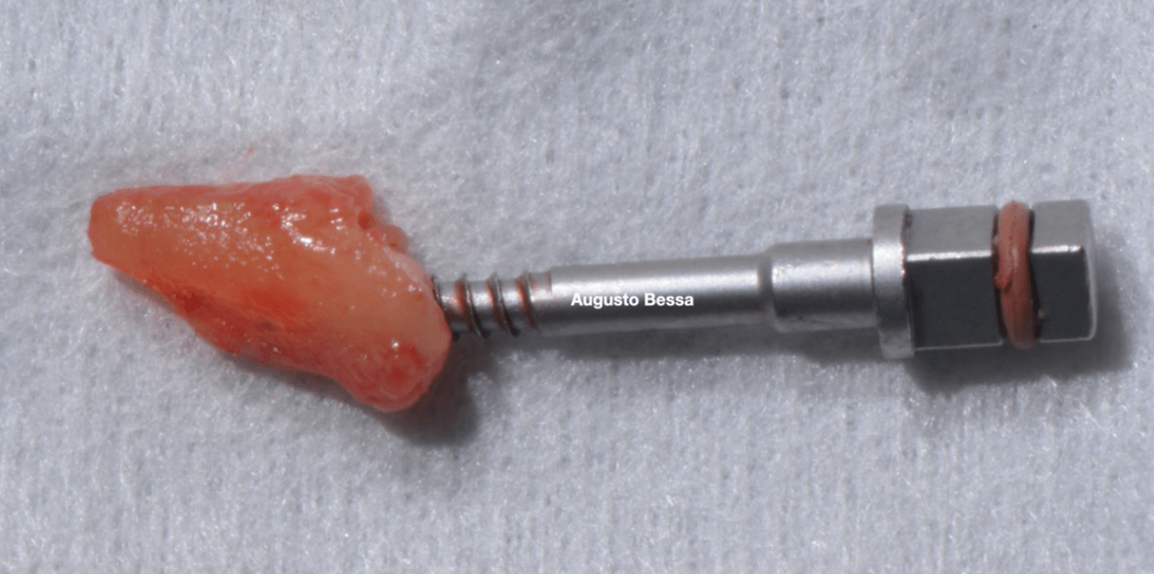
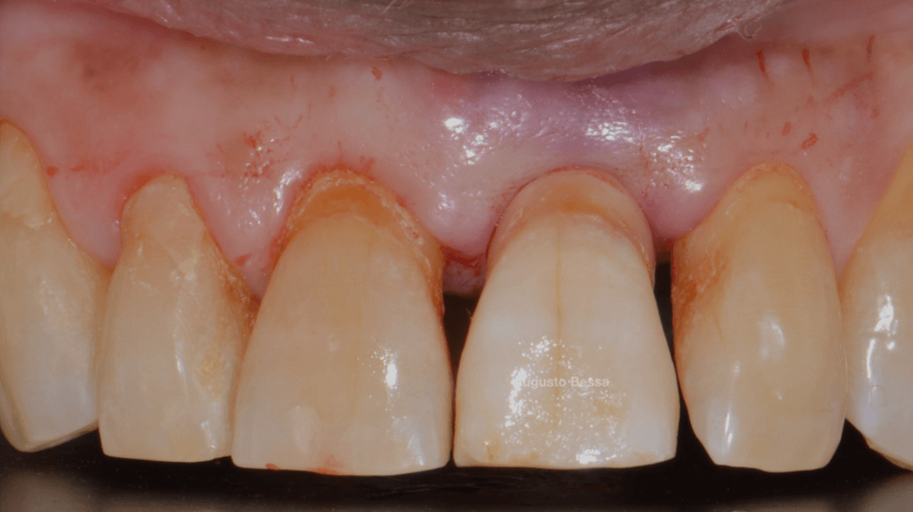
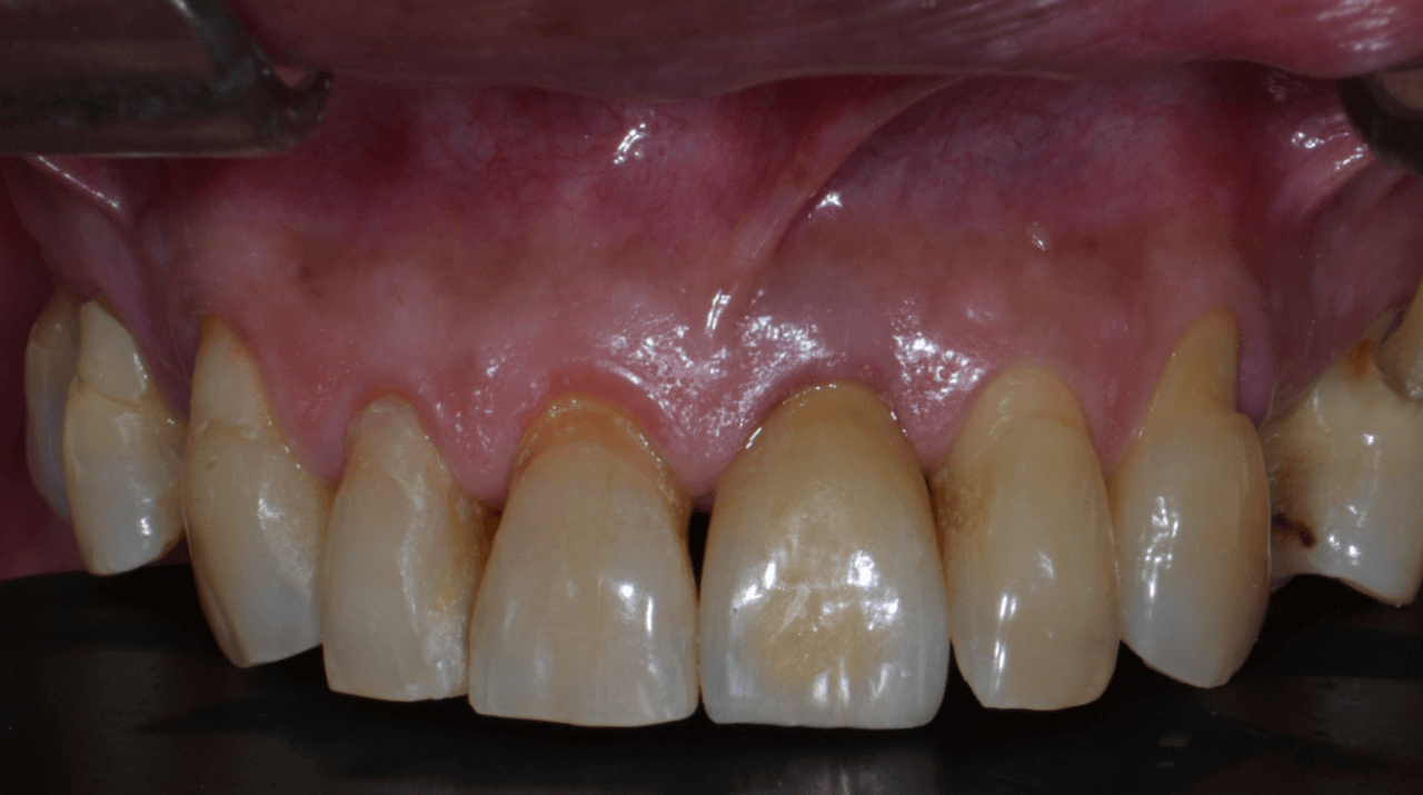
Male patient, 61 years old, went to the private dental clinic after suffering trauma to dental element 21 (Fig. 1). The clinical examination showed mobility of the crown, and the horizontal fracture was confirmed by the periapical radiographic examination (Figs. 2 and 3), and surgical removal of the root and rehabilitation with an implant was indicated. After the tomographic examination, the case was built virtually in the 3Shape Implant Studio program, and the planning for the atraumatic exodontia of the root remains was carried out (Fig. 4), aiming to preserve the sorrounding bone structure and facilitate the surgery for installing the immediate implant. The chosen implant system was Arcsys by FGM.
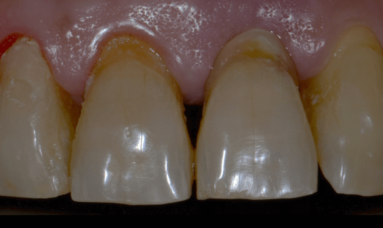
Figura 1 Exame clinico inicial - Treatment of a fracture on an upper central incisor with rehabilitation through immediate implant carried out with a virtual surgical guide: a clinical case report
Fig. 1- Initial clinical examination.
Figura 2 Fratura do elemento dentario do 21 co - Treatment of a fracture on an upper central incisor with rehabilitation through immediate implant carried out with a virtual surgical guide: a clinical case report
Fig. 2- Fracture of the dental element of tooth 21 confirmed through a periapical x-ray.
Figura 3 Remocao da coroa fragmentada - Treatment of a fracture on an upper central incisor with rehabilitation through immediate implant carried out with a virtual surgical guide: a clinical case report
Fig. 3- Removal of the fragmented crown.
Figura 4 Extracao atraumatica do elemento dent - Treatment of a fracture on an upper central incisor with rehabilitation through immediate implant carried out with a virtual surgical guide: a clinical case report
Fig. 4- Atraumatic extraction of the dental element of tooth 21.

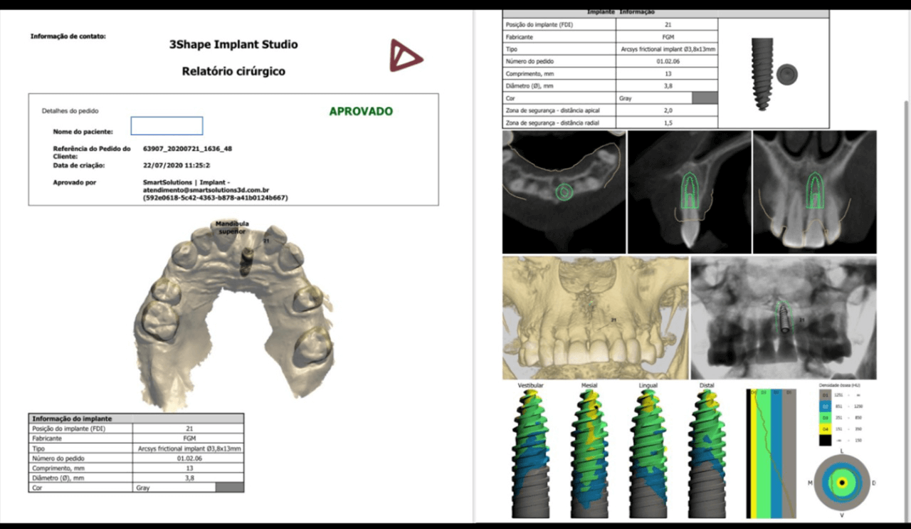
Before the surgery, the virtual surgical guide was planned, joining the STL file of the scanning to the DICOM file of the tomography in a specific planning software (Figs. 5 and 6).
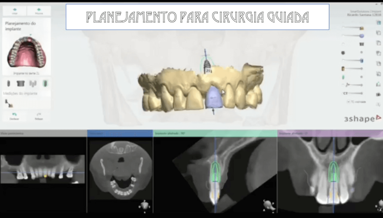
Figura 5 Planejamento para guia cirurgico 3Sha - Treatment of a fracture on an upper central incisor with rehabilitation through immediate implant carried out with a virtual surgical guide: a clinical case report
Fig. 5- Planning for the 3Shape surgical guide.
Figura 6 Relatorio cirurgico aprovado - Treatment of a fracture on an upper central incisor with rehabilitation through immediate implant carried out with a virtual surgical guide: a clinical case report
Fig. 6- Approved surgical report.

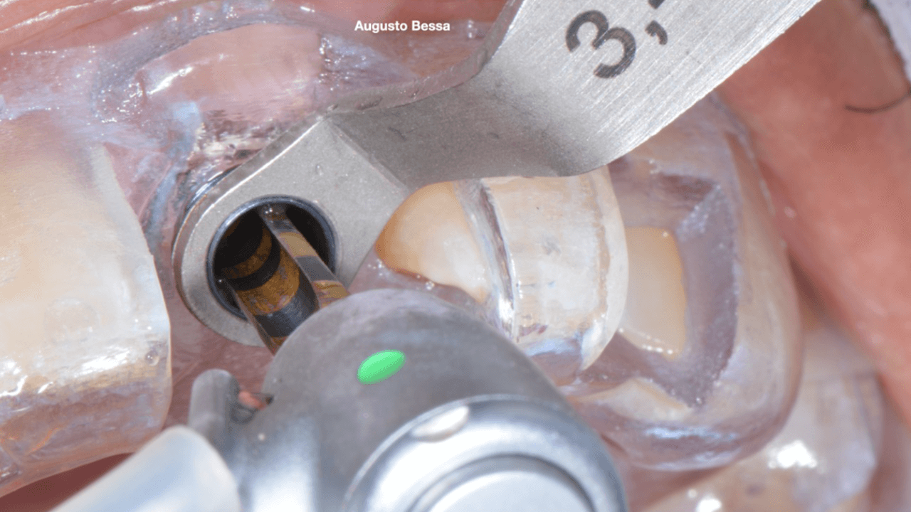
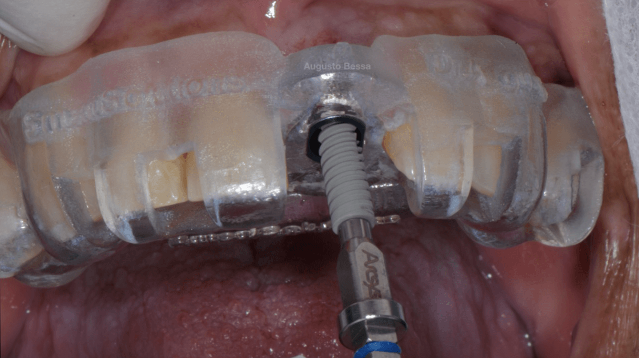
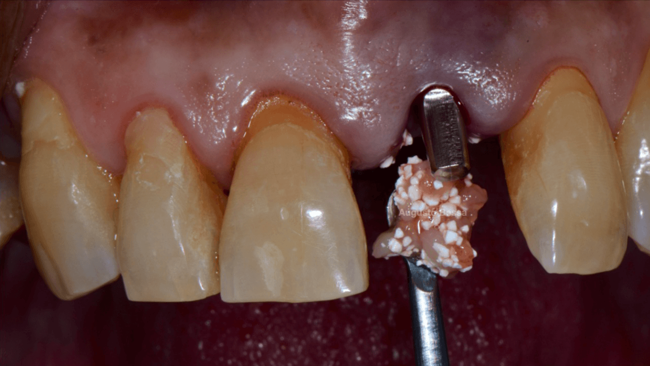
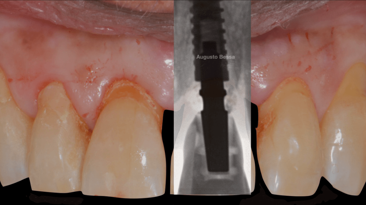
With the aid of the surgical guide printed from the virtual planning (Fig. 7), the surgical instrumentation was executed to install a 3.8 mm x 13 mm implant (Fig. 8). The implant was installed (Fig. 9) and anchored to the alveolus. A 3 mm x 6 mm Arcsys prosthetic abutment with a 4.5 mm transmucosal portion was activated according to the protocol recommended by the manufacturer. The filling with the Nanosynt bone graft with granulation from 500 µm to 1000 µm was performed (Fig. 10), associated with the chopped L-PRF fibrin.

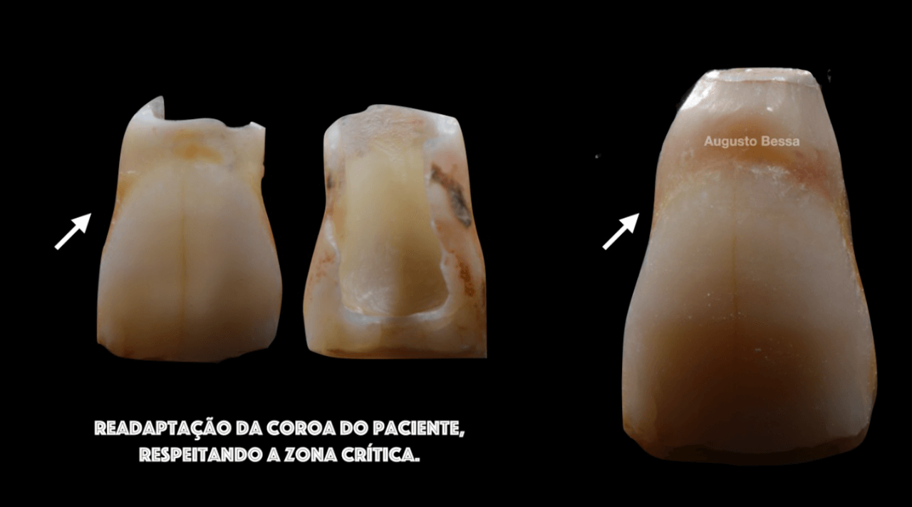
A provisional crown was made immediately through the fractured crown, respecting the critical zone (Fig. 11). After polishing and finishing, it was cemented to the abutment with provisional cement (Figs. 12 and 13).
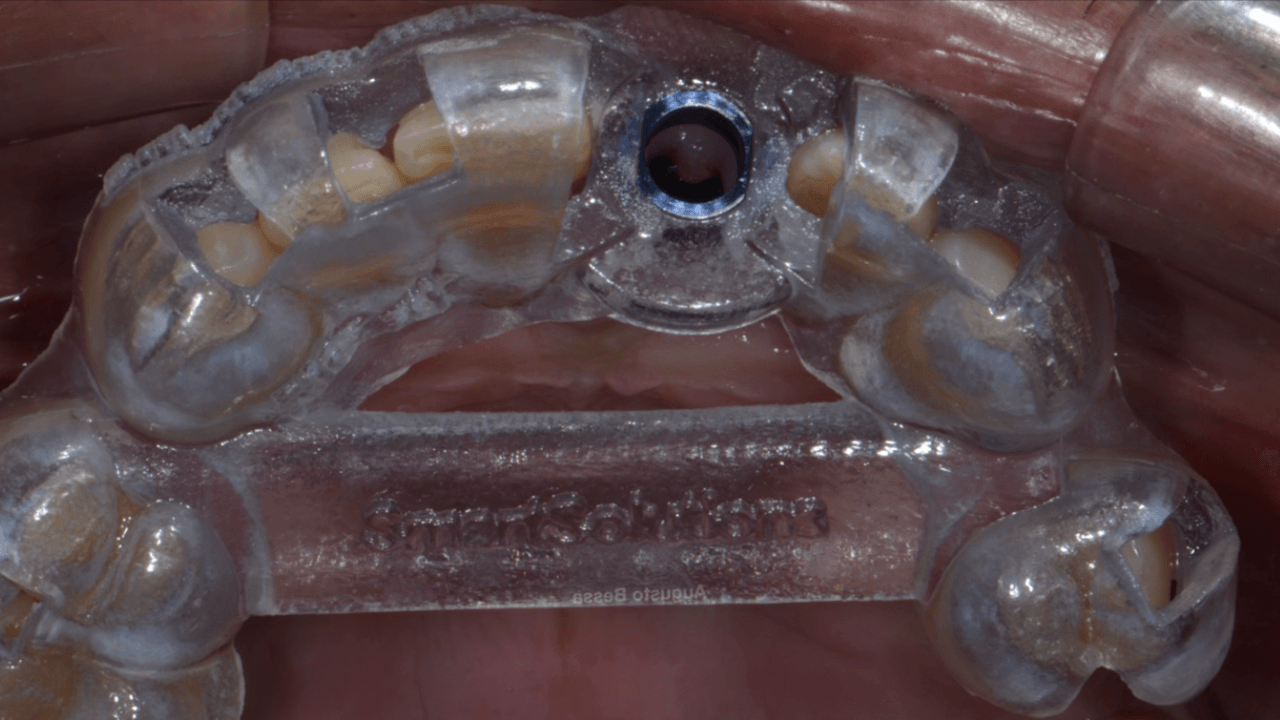
Figura 7 Guia cirurgico - Treatment of a fracture on an upper central incisor with rehabilitation through immediate implant carried out with a virtual surgical guide: a clinical case report
Fig. 7- Surgical guide.
Figura 8 Broca cirurgica de perfuracao de acor - Treatment of a fracture on an upper central incisor with rehabilitation through immediate implant carried out with a virtual surgical guide: a clinical case report
Fig. 8- Surgical drilling bit following the guide.
Figura 9 Instalacao do implante - Treatment of a fracture on an upper central incisor with rehabilitation through immediate implant carried out with a virtual surgical guide: a clinical case report
Fig. 9- Installation of the implant.
Figura 10 Preenchimento com Nanosynt e L PRF - Treatment of a fracture on an upper central incisor with rehabilitation through immediate implant carried out with a virtual surgical guide: a clinical case report
Fig. 10- Filling with Nanosynt and L-PRF
Figura 11 Provisoria confeccionada - Treatment of a fracture on an upper central incisor with rehabilitation through immediate implant carried out with a virtual surgical guide: a clinical case report
Fig. 11- Provisional crown made.
Figura 12 Coroa provisoria cimentada - Treatment of a fracture on an upper central incisor with rehabilitation through immediate implant carried out with a virtual surgical guide: a clinical case report
Fig. 12- Provisional crown cemented.
Figura 13 Implante instalado e coroa provisori - Treatment of a fracture on an upper central incisor with rehabilitation through immediate implant carried out with a virtual surgical guide: a clinical case report
Fig. 13- Implant installed and provisional crown captured.

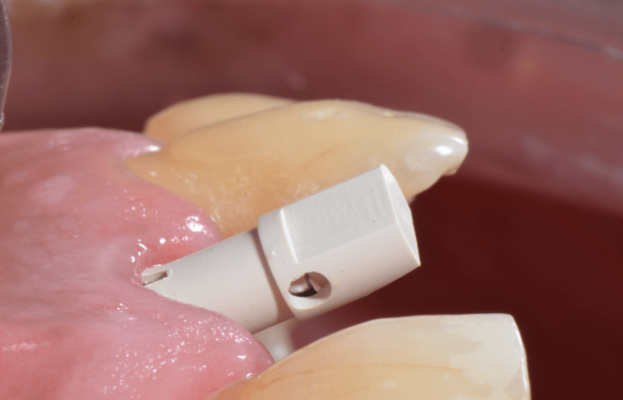
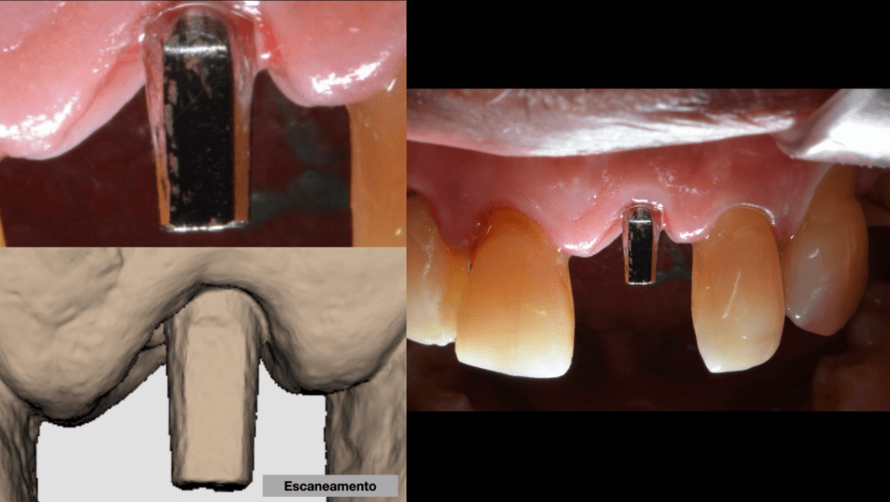
After 4 months of waiting for the bone remodeling of the alveolus and the osseointegration of the implant, we proceeded to execute the connective tissue graft, improving the pink peri-implant esthetics. After this step healed (Fig. 14), the prosthetic rehabilitation was planned, also in the digital workflow. For such, the Scan Body for abutments was used, designed with an inspection window to facilitate its visualization on the prosthetic component (Figs. 15 and 16). The selection and photography of the colors were performed to send to the laboratory (Fig. 17).
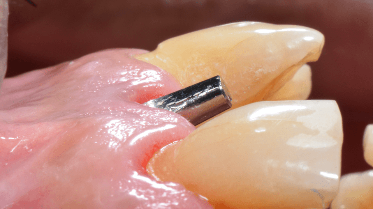
Figura 14 Estetica Periimplantar apos a cicatr - Treatment of a fracture on an upper central incisor with rehabilitation through immediate implant carried out with a virtual surgical guide: a clinical case report
Fig. 14 – Peri-implant esthetic after the healing of the connective tissue graft.
Figura 15 Janela de inspecao do Scan body do m - Treatment of a fracture on an upper central incisor with rehabilitation through immediate implant carried out with a virtual surgical guide: a clinical case report
Fig. 15 – Inspection window of the Scan body abutment.
Figura 16 Escaneamento do nunhao protetico - Treatment of a fracture on an upper central incisor with rehabilitation through immediate implant carried out with a virtual surgical guide: a clinical case report
Fig. 16- Scanning of the prosthetic abutment.
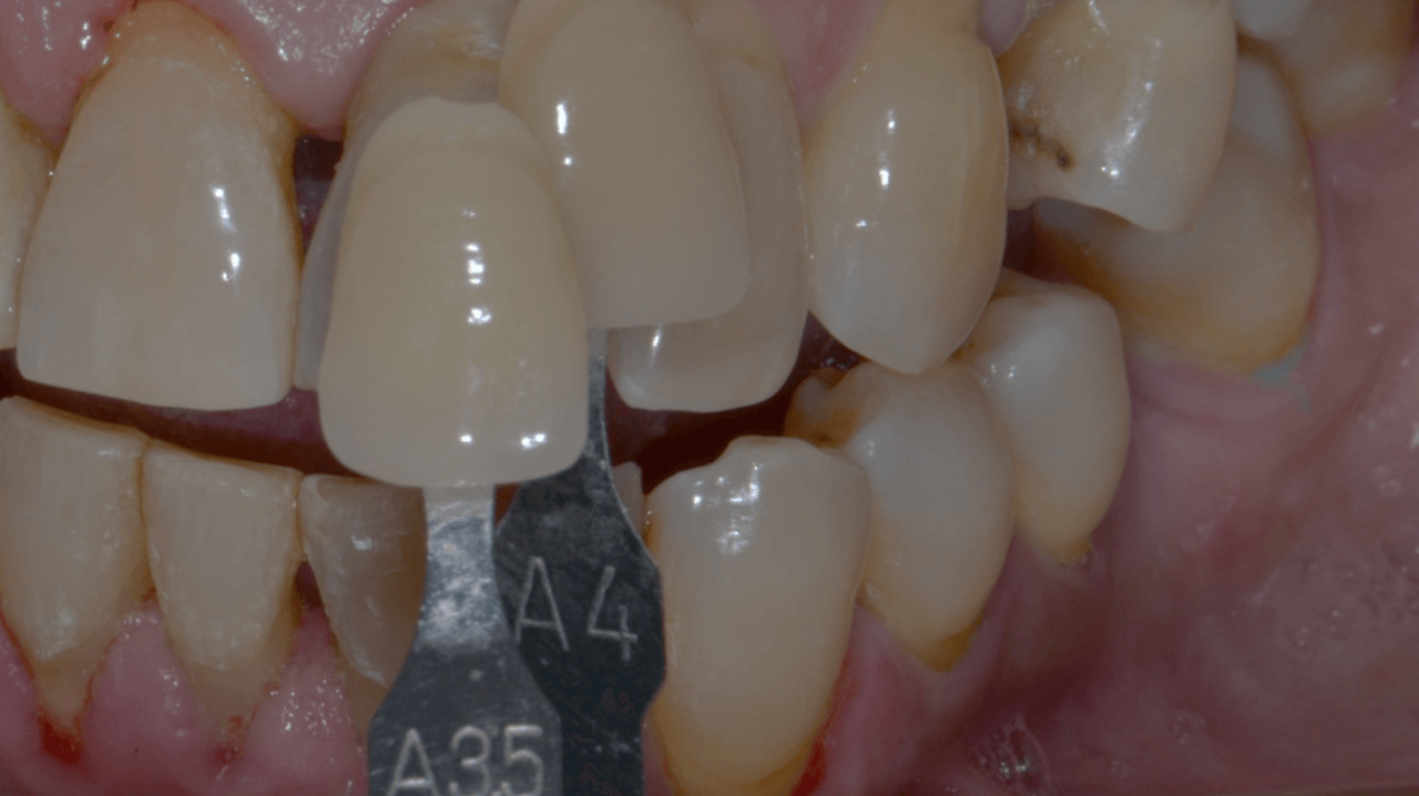
Figura 17 Selecao de cores - Treatment of a fracture on an upper central incisor with rehabilitation through immediate implant carried out with a virtual surgical guide: a clinical case report
Fig. 17- Shade selection.

Considering that the library of Arcsys implants and components is available in the 3Shape software, the CAD operator planned, designed, and milled a zirconia cast and performed the stratification of the ceramics. Upon removing the provisional crown, the healed emergence profile with the tissue positioned favorably was observed (Fig. 18).
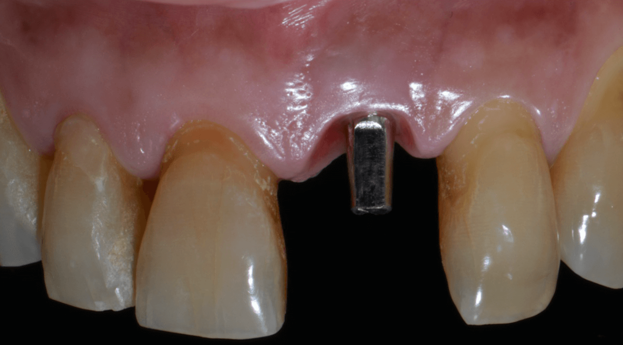
Figura 18 Perfil de emergencia - Treatment of a fracture on an upper central incisor with rehabilitation through immediate implant carried out with a virtual surgical guide: a clinical case report
Fig. 18 – Emergence profile.

The case was finalized with a perfect adaptation (Fig. 19), and the definitive prosthetic cementation was performed (Fig. 20). The replacement of dental elements in the anterior region using the digital flow results in more significant swiftness in the surgical act and facilitates the clinic’s day-to-day, increasing the comfort, precision, and quality of the results.
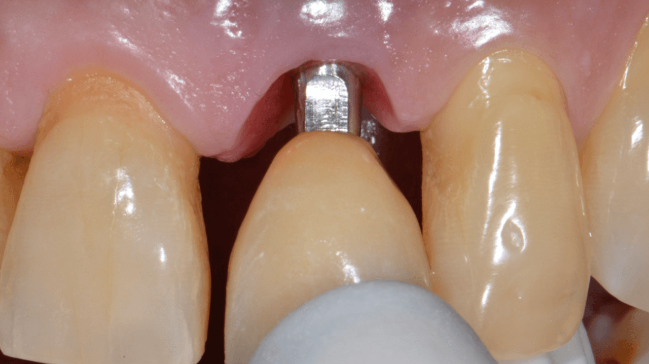
Figura 19 Prova da coroa definitiva. Observa s - Treatment of a fracture on an upper central incisor with rehabilitation through immediate implant carried out with a virtual surgical guide: a clinical case report
Fig. 19 – Definitive crown tryout. the fit between the crown and the tissue.
Figura 20 Cimentacao final - Treatment of a fracture on an upper central incisor with rehabilitation through immediate implant carried out with a virtual surgical guide: a clinical case report
Fig. 20 – Final cementation.

References

  1. Catálogo de produtos da FGM Dental Group;
  2. Regeneração Óssea Guiada na Implantodontia
    Daniel Buser – Quintessence Editora – 2010
  3. Regeneração Guiada Tecidual
    Mauro Cruz – Editora Santos – 2006
  4. Enxertos ósseos em Implantodontia
    Jean-François Tulasne e Andreáni – Quintessence Editora – 2010
  5. Enxertos Ósseos em Implantodontia
    Reanato Mazzonetto et al. – Napoleão Editora – 2012
  6. Transplantes Ósseos na Implantodontia
    André Antonio Pelegrine et al. – Napoleão Editora – 2008
  7. Catálogo de produtos da FGM http://www.fgm.ind.br/site/produtos/implantesbiomateriais/substituicao-ossea-nanosynt/
  8. Choukroun, J. (2001) Une opportunit_e en paroimplantologie: le PRF. Implantodontie, 42, 55– 62. French.
  9. Pan J, Xu Q, Hou J, Wu Y, Liu Y, Li R, Pan Y, Zhang D. Effect of platelet-rich fibrin on alveolar ridge preservation: A systematic review. J Am Dent Assoc 2019; 150:766-78. doi: 10.1016/j.adaj.2019.04.025.

Email
LinkedIn
Telegram
Facebook

Most recent posts

plugins premium WordPress

Selecione seu idioma

Select your language

Shop FGM Implants

Componentes Arcsys, Vezza e Fluxo Digital até 60% OFF, promoções em Brocas, kit cirúrgicos e muito mais.