![Reconstrucoes-osseas-e-implantacoes-com-guia-prototipada-em-areas-de-agenesia-1[1]](https://fgmdentalgroup.com/wp-content/uploads/2022/11/Reconstrucoes-osseas-e-implantacoes-com-guia-prototipada-em-areas-de-agenesia-11.jpg "Reconstrucoes osseas e implantacoes com guia prototipada em areas de agenesia 11 - Bone reconstruction and implantations with prototyped guide in agenesia areas")
Authors: Dr. Rafael Cury Cecato, Dr. Thiago Roberto Gemeli and Dr. Ricardo de Souza Magini
29-year-old female patient.
CHIEF COMPLAINT
Functional and esthetic dissatisfaction with the partial removable prosthesis, responsible for the replacement of elements 12 and 22.
INITIAL EVALUATION
After anamnesis the patient was submitted to clinical radiographic assessment to ratify the agenesia of elements 12 and 22. Besides the dental elements, the exams revealed the bone volume deficiency in those regions, as well as a limited space in the mesial-distal direction (between 13/11 and 21/23) of the referred elements.
TREATMENT CARRIED OUT
The present report demonstrates, step-by-step, the surgical procedure carried out in a 29-year-old female patient, with congenital analgesia of the upper lateral incisors. Clinical and image exams revealed the need for the reconstruction and rehabilitation of the regions of teeth 12 and 22, according to figures 1 to 5.


INTRODUCTION
The rehabilitation with stability of the peri-implant tissues in borderline cases is always a challenge. The predictability of results requires the use of materials, techniques and devices that support the requirements associated to the health, function and esthetics of each case, which demands not only the use of contemporary technologies but, above all, extreme dedication and respect to the surgical-prosthetic plan.
STEP-BY-STEP
The initial proposal, with orthodontic approach for the correction of the dental classes, medium line and interradicular distances between the central incisors and canines was not accepted by the patient. The option was then to initiate the reconstruction and follow with the rehabilitation.
The re-planning considered the horizontal volumetric increase of the bone plates, followed by the implantations assisted by prototyped surgical guides and prosthetic rehabilitation with ceramic crowns. The problems associated with the congenital absence of the dental elements involved not only those affected by the narrowing of the ridges but, specially, incurred in a risk for damage to the surrounding radicular structures. That condition, allied to other advantages, justified the surgical approach assisted by surgical guides since the ideal tridimensional positioning predictability of the implants allowed through this technique is proved to be greater.
Fundamentally, the guided surgery offers greater operational safety and may bring indirect benefits, decreasing the time in the office and morbidity. To decrease the problems that interested the patient, the treatment was developed in three different phases:
- Horizontal bone tissue increases with synthetic biphasic ceramics.
- Implantation of implants in the regions of elements 12 and 22 with prototyped guide.
- Provisioning for tissue conditioning and finalization with total crowns in porcelain.
In phase I the synthetic biphasic ceramic (Nanosynt – FGM) was associated to platelet aggregates in order to offer mechanical stability and greater metabolic dynamism to the region to be regenerated. The choice for this biomaterial was in function of its physical-chemical properties related to the osteoblastic activity and also for presenting excellent osteoconductive standards, promoting cell stabilization and colonization over itself, optimizing the tissue response.


Indifferent to the greater thickening of the ridge obtained, the intraradicular spaces corresponding to the agenesia remained narrow. For that, CT scans and scanning will allow the overlaying and manipulation of those files in specific software, with the purpose of allowing for the impression of a prototyped guide which will help the installation of the implants, guiding them, in the ideal tridimensional position.

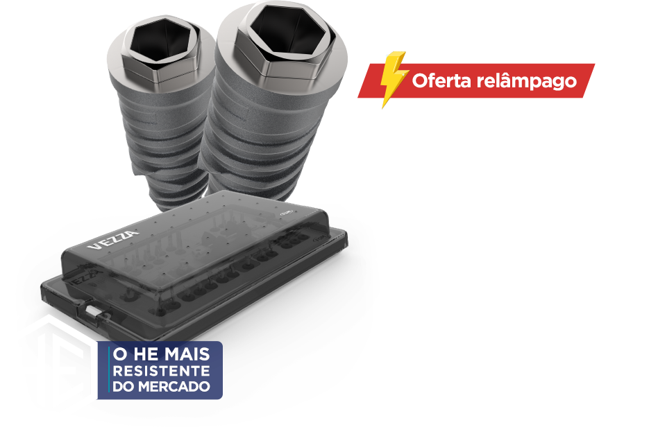
Although the technique may be executed through a number of systems, its results become even more interesting with the Arcsys (FGM) system. The particularity of the refinement of the angle in an infinite number of degrees, potentializes even more the reach of the results, contributing to a better biomechanical and esthetic performance.
After the approval of the positioning of the implants by the professional, a prototyped guide is digitally modeled and printed, in order to be used in the surgery.


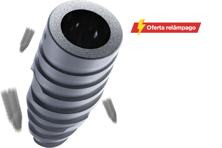
In Phase II, destined to the implantations, the guide is attached to the dental arch and its stability is checked. The design of the AGS drills (FGM) allow for a single perforation, minimizing even more the procedure times and contributing significantly to the agility of the procedure. According to the recommendations of the manufacturer, in face of the diameter of 3.3mm of the selected implant (fig. 18), the perforations were carried out just with the 2.4mm drill. The drill guides allowed for precision throughout the instrumentation (fig. 19).


The implants were installed with the help of the installation driver. It has two functions, because besides allowing for the capture of the implant, it has an anti-locking mechanism Before activating the prosthetic component over the implant, as previously planned, the customization of its angulation was promoted in the clinic, by the professional and enough resistance to be used also as a final installation drive with torque wrench, with safety even when expressive torques are needed (figs. 20 to 22).


Before activating the prosthetic component over the implant, as previously planned, the customization of its angulation was promoted in the clinic, by the professional and enough resistance to be used also as a final installation drive with torque wrench, with safety even when expressive torques are needed (figs. 20 to 22). carrying out the treatment. The customization mentioned is obtained with the use of a template denominated angle referrer (figs. 24 to 27).


After the activation of the intermediate pieces and provisioning. A 90 day period was respected.